昨年度はTARA用実験ステーション建設を最重要課題として取り組み、計画 通り、平成8年5月より運転を開始した、この経過報告を始め、ソフトウェアー、 ネットワーク、周辺装置等の整備状況並びに、本プロジェクトとしての論文を報 告する。
I. TARA用実験ステーション
先回も述べたようにビームラインの建設はPFの渡辺信久氏(本誌Vol.1,No.2. 87頁)、ハッチは鈴木守氏(本誌Vo1.2,No.1,107頁)、装置関係は坂部知平 (本誌Vol.1,No.129頁)等が、それぞれ中心となって進めれられた(本誌Vo.2, No.1 95頁)。
1.ビームライン
変更電磁石から放射された放射光はまずメインシャッター(MS)やブランチ ビームラインにビームを分岐するスリット及びブランチビームラインシャッター (BBS)、その他多くの安全装置等を備えた機関チャンネルと総称される部分 を通過し光源の壁の外側にある実験ホールヘと導かれる。ここから後が我々が製 作を受け持つ部分である。これはビームライン、ハッチ、測定器とに大別される。 ビームラインは放射光を導くパイプ、高調波を除去し且つ垂直方向にビームを集 光するためのミラー、放射光の確認のための蛍光板やその覗き窓、ダウンストリ ームシャッター(DSS)等が目立つが実はその他、スリット、真空維持のため に各所に設けられたベリリウム窓と真空ポンプが重要な役を演じ、更に各所にイ ンターロックが設けられ安全運転を監視している。
ビームラインの組立が終了し最初に遭遇するのはビームラインの真空が規定ま で上がるかどうかである。高真空を出すのは職人芸でネジの締め方一つで真空が 破れると言われている。新しい金属はまるでスポンジのようにガスを放出するの で安定した高真空を得るのには各所に熱をかけ焼き出しを行うが、それでも時間 の掛かる仕事である。真空が得られるとインターロックのテストが行われる。こ のテストは3月21日に行われた。しかしこれですぐビームが使えるわけではな い。放射光を通すとダクト内の各所からガスが放出され、真空度が下がりインタ ーロックが作動してメインシャッターが閉じてしまう。そこで、光源の電流を極 く低いところから徐々に上げてもらい、ビームラインの焼き出しを行う必要があ る。この作業が4月1日に行われた。この日、光源の電流が上がるに従い、放射 光の漏洩がないか注意深く測定され、漏洩箇所が見付かるとそこに鉛板を張る作 業が行われた。一般的に言ってフランジ(継ぎ手)付近に漏洩が起こりやすい、 単純な作業であるがこれが意外に難しく、下手をすると巻いた後鉛板が皺くちゃ になる。
2.カメラ
4月26日にTARA用カメラの設置を行った。幅2.68mしかないハッチ にべ一スが2.44mの幅を持つ数トンのカメラを設置し、しかもビームの中心 とカメラの光軸の誤差5mm以内、ビームと光軸の平行からのズレが3度以内と いう厳しい条件での設置は大変な作業であった。まずミラーは集光度良く合格、 次にダイレクトビームとカメラの光軸を合わせ、非対称カット(α=7.8度) 三角ベントSi(111)を取り付け、全波長領域でX線とカメラの光軸を一致 させる必要がある。これは極めて精度を要する作業である。また、カメラの精度 のテストも必要である。もっとも大切なのは試料の回転軸と光軸の交わりである。 測定の結果70μm程の不一致が観測され、修正を行った。このほか2θアーム の動き及び光軸調整用の水平面内角度調整機構の動きがスムーズさを欠いている ことが判明した。工場まで送り返して直すとなると春期の利用開始は全く不可能 になる。このままでも、PFスタフ(渡辺信久、鈴木守両氏)が調整すれば波長 を変えられるが共同利用者が調整することは少し難しい。結論として、春期は波 長を1.OAに固定し、ビームタイム終了後に修理することにした。もう一点 θの回転をパルス当たりO.O02としたが、実はこれではMAD法の際、試料 結晶のEXAFSを測定し正確に波長を合わすには荒すぎることが分かりパルス 当たりの動きを0.0002度に改良した。このほか多くのテストを行い、5月 22日にテスト期間としながらも一応利用者にオープンした。
3.思いがけない出来事
利用者にオーブンして間もなくハッチの温度を4度に下げると光が来なくなる と言う苦情が起こった。これはBL6Aでは起こったことのない現象で、一瞬驚 いたが、鉄の膨張係数を基に計算すると1度当たり約8μmの伸縮があることが 分かった。即ち、15度近い温度変化に対し約120μmの収縮が起こり、TA RAビームラインのミラーによる集光が良好なため、カメラの光軸がビームから 外れた訳である。BL6Aの場合は集光を甘くしてあるため、この現象がはっき り現れなかったのである。つまりミラーの集光が良いため、最初の予想を上回る、 BL6Aの約2倍の輝度を得ることが出来た代償である。この問題はハッチの温 度を7度以上変えた場合、3時間ほど経ってから光軸合わせを行えば済むことで あるからなんらの処置も行わなかった。
4.大型IP読取装置
犬型IP読取装置には多くの問題点があった。最も困ったのはIPの排出が巧 く行かず、IPを傷つけるトラブルが幾度も発生し、1枚25万円のIPが一瞬 にして傷物になった。泣いても泣ききれない!これはセンサーを増し、各所のタ イミングを取り直し可成り良くなったがそれでも時折引っかかった。結局出口に IPの曲がりを強制的に減らす押さえを置くことで何とか止めることが出来た。 装置の立ち上げというのはこの様な問題が常に生じるもので、今回が特別ではな い。この様な葛藤の内に時間が経ち、6月の半ばになってやっと安定した。
今もっともネックになっているのは大型IPの読取に1枚当たり10分強かか ること及び電子計算機のメディアであるDATにデータを落とすのに可成りの時 間が掛かることである。後者については後述するプレハブに高速でデータを転送 しプレハブ内でDATに落とすことを計画している。詳しくは六号に名古屋大学 の佐々木教祐氏が記載しているので参照されたい。
5.TARA用実験ステーションの特徴
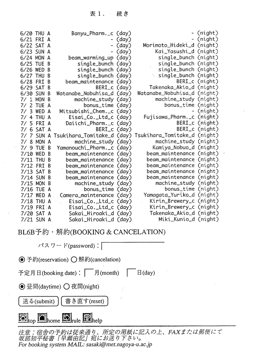
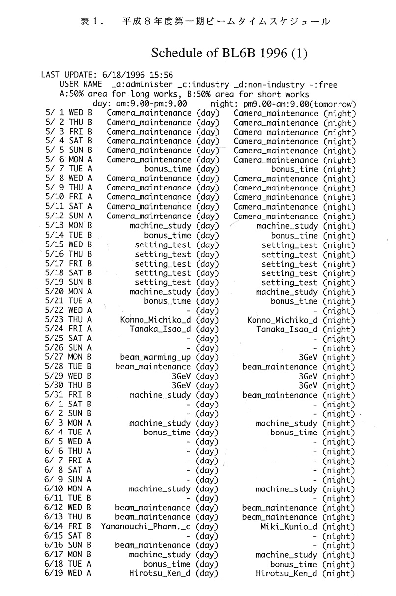
以上問題点のみ列挙したが、実は、輝度が大きく、カメラが使い易く、大型の IPが使用できしかも精度も高いこと等により、6月半ばより次第に好評の声が 聞こえるようになって、ホッと胸をなで下ろしている。妻1に本年第一期のスケ ジュール表を示す。良いとなるとたちまちスケジュール表が埋まり、嬉しい悲鳴 に変わった。良い点を次に示す。
最小カメラ長;575.7±O.07mm
最大カメラ長;967.9±O.14mm
Ⅱ.電子計算機及びソフト
1.実験ホールの近傍に設置したプレハブと実験ステーション間を光ファイバー ケーブルで結ぶ予定である。これら電子計算機及びそのネットワークについては 6号に掲載された佐々木教祐氏の記事を参照されたい。
2.WWWに我々のホームページがあり(本誌Vol.1,No.296頁)ここにTARAで 使用できるソフトウェアーについて詳しい記事が載っているが気付いていない人 が多いようなので少し紹介する。これはTARAのビームタイム予約と電子計算 機の予約のためのもので佐々木教祐氏が開設し管理している。これを開くとまず 表2の画面が現れ、「TARA COMPUTERの使用予約」をクリックすると表3が表示さ れる。更に「各コンピュータで使用出来るソフトについての説明」をクリックす ると表4が表示され、現在TARAとして使用できるソフトウェアーを知ること が出来る。未だ続くが切りがないので中止する。現在これらソフトの管理も全て 佐々木教祐氏が行っている。
3.上記ソフト中Xsight及びQUANTA/CHARMmで代表されるプログラムパッケージ はそれぞれ㈱菱化システム及びCTCラボラトリーから購入したもので、表5及 び表6に示す多くのプログラムを包含している。尚、購入にあたりこれらは高価 なプログラムパケージであるためソフトウェアー検討会議にて検討を行った。ソ フトウェア検討会議については本誌Vol.2,No.1 104及び109~112頁に記載されて いる。
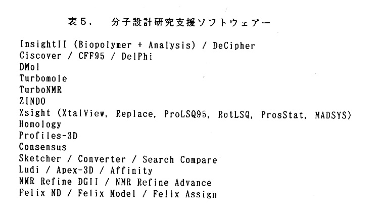
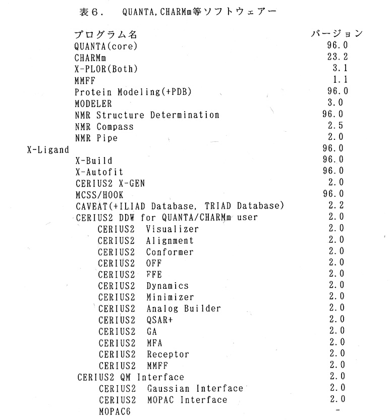
4.これらのソフトは構造解析以外のものも含まれている。使用に当たって分か らないことは業者が積極的に対応してくれることになっている、そして講習会も 場合によっては、企業或いは研究室単位でも開いてくれる約束である。年に数回 の講習会をTARAで開催することを積極的にTARAが引き受ける契約になっ ている。この場合TARA以外の人も自由に参加出来る。尚、表5及び表6に示 すプログラムは「同一のプログラムを同時に2カ所以上」で使用することは出来ない。将来使用頻度が多くなった場合は契約の変更をする必要がある。またこれ らのソフトを使用した研究成果を公表する場合「Mo1ecular Simulations Inc.」の 名称を出来る限り掲載すること」になっている。
㈱菱化システム 担当者・氏名 狩野敦氏
Te1.0473 80 1231
Fax.0473 80 1239
CTCラボラトリーシステムズ㈱ 担当者・氏名 松永公人
Te1. 03 3419 9171
Fax. 03 3419 9179
e-mai1 matsu@ctcls.co.jp
5. 萬有製薬㈱の三浦圭子さんが本号に「Xsightの案内 その1」と題し、彼女 の意見を交えながら具体的に使用方法を説明した案内記載が掲載されているので 参照されたい。
Ⅲ. 仮設型収納施設(プレハブ)
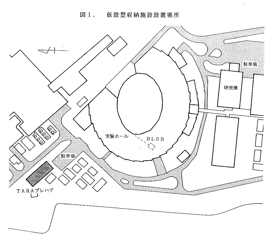
TARAプロジェクトは筑波大学に属するものであるが我々のプロジェクトは 放射光を使用するためそれに関連する設備は高エネルギー物理学研究所(KEK) 内に設置すると大変効率よく研究が進む。その代表的なものがTARA用実験ス テーションである。しかし、実験ステーションは単独に存在して効率を上げ得る ものではなく、そのまわりに数多くの電子計算機、結晶準備室、その他装置の立 ち上げに必要な場所など多くの機器や場所が必要である。幸い図1に示す場所に KEKより仮設型収納施設設置場所として240m2の土地の借用が認められた。
当初2階を有する収納施設を予定していたが色々な困難があり今年度はこの場 所に3個の収容施設を設置する計画を進めている。既に最初の収容施設は設置を 終了した。ここは数多くの電子計算機を収納し、実験ステーションとの問を光り ケーブルで繋ぎ、データの処理を行うためのものである。名古屋大学の佐々木教 祐氏が6号に詳細を書いているので参照されたい。残り2つの収容施設に付いて も現在購入,を検討し、出来ることなら次回のビームタイム(11月)に間に合わ せたいと思っている。
KEK施設部の協力で、50KW及び25KW(動力用)等2個のトランスを PF内の電源室にに設置することが認められ、更に収容施設近傍までの電力工事、 及び給排水等が可能になった。
Ⅳ. 単結晶構造解析用迅速X線解析装置(自動回折計)
オックスフォード社製低温窒素気流吹付型試料冷却装置をTARAとして1 台購入した。これはPFでBL6A用及びBL18B用として購入したものと同 型である。即ち合計3台実験ホール内にあるので都合し合えば余裕を持って使用 できる。
Dynapro.801を購入した。これは溶液散乱を測定し結晶化条件をサーヴェイす るための装置で温度制御も出来るものである。PF研究棟1階の低温室の隣の部 屋(製氷器や、インキュベータがある部屋)に設置されている。既に使用経験の ある萬有製薬㈱の三浦圭子さんにテストを依頼した結果使い易く良好との報告が あった。
Ⅸ. ビームラインアシスタント及び保守要員
平成8年度前期のビームラインアシスタントの希望者は7人である。2人制を 取る予定であったが、今回は希望者が意外に少なく事実上1人で頑張ってもらっ た。結果として、1人では負担が多く極めて気の毒であった。来期は2人制が実 行出来るよう努力する。
平成8年度前期のビームラインアシスタントの方々の名簿を表7に示す。
表7. 平成8年度ビームアシスタント名簿
|
氏名 |
学年 |
所属 |
指導教官名 |
|---|---|---|---|
|
松本拓男 |
M2 |
長岡科学技術大学 |
三井幸雄 |
|
岡田健吾 |
D3 |
大阪市立大学 |
広津建 |
|
富長隆裕 |
D2 |
北海道大学 |
田中勲 |
|
齋藤純一 |
D2 |
京都大学 |
三木邦夫 |
|
菅原光明 |
D2 |
京都大学 |
三木邦夫 |
|
藤掛真広 |
M2 |
名古屋大学 |
山根隆 |
|
杉浦郁子 |
D2 |
お茶の水女子大学 |
今野美智子 |
今春よりPFはTARA実験ステーションを含む、生物関連機器のための保守
要員として、理学電機㈱より出向と言う形式で技術員を1人雇用した(本誌Vo1.
2,N0.1106頁)。
この方の名前は中丸幸雄氏、電気科出身でサービス(機器の出張修理)を主と
して来られたが、近年設計1課、電気1課製品開発部等を経て今回PFに出向し
て来られた。着任されたのが丁度TARA実験ステーションの立ち上げ時期でも
あり、大変活躍された。
Ⅶ. 各種委員会報告
1. 行事委員会報告
6月21日、筑波大学に於いて第2回パネルディスカッションを開催した。
テーマは「電子密度の改良」で、千田俊哉(長岡技科大)、宮野雅司(JT)、
山下栄樹(仮大蛋白砂)、鈴木津巨(名大工学部)の諸氏が話題提供を行った後、
参加者52名で討議した。
2. 編集委員会
第4回編集委員会を9月11日18時より筑波大学にて開催した。委員長より
構造生物Vol.2,No.2の原稿が殆ど集まったとの報告があり、印刷、配布など、具
体的な決定がなされた。また、次号は総会後になるので、総会の記事及び学術記
事・研究室紹介等の執筆者の人選などについての意見交換が行われた。
Ⅷ. 放射線業務従事者登録更新時教育訓練の実施
放射光実験施設を利用するためには放射線常務従事者登録が必要である。本プ ロジェクトは人数が多いため、全学放射線管理委員会委員長の承認の下に、筑波 大学アイソトープセンター(放射線総括管理者池田龍一教授)の御厚意で本プロ ジェクトのために平成8年5月13日(月)及び6月24日(月)の2度に渡り、 放射線業務従事者登録更新時教育訓練を行って頂いた。参加者はそれぞれ54及 び15人であった。講師はアイソトープセンター放射線取扱主任者海老原寛先生、 アイソトープセンターの世話人は松尾先生、田倉先生、西山専門職員、傳田主任 の4人であった。その後1名の受講者が出たが、それは全学の教育日に出席して 頂いた。その結果希望者全員支障無くTARA実験ステーションを利用できた。
Ⅸ. 業績紹介
論文中にTARAのメンバー或いはTARAに謝意等を表明され、送付されて きた論文のリストを記載する。いずれ論文数が増えてきたら独立に欄を設けるべ きであるが、まだ始まったばかりで数も少ないのでここにまとめる。
1.Hartmut Michel、岩田想グループの論文
論文1
Structure at 2.8A resolution of cytochrome
c oxidase from Paracoccus
denitrificans. So lwata, Christian Ostermeier,
Bernd Ludwig* & Hartmut
Michel, Nature Vol 376・24 August 1995, 660-669.
Max-Plank-Institute fur Biophysik, Heinrich-Hoffmann-Strasse
7, D-60528
Frankfurt/M. , Germany. *Johann-Wolfgang-Goethe
Universi tat, Biozentrum,
Institut fur Biochemie, Molekulare Genetik,
Marie-Curie-Strasse 9, D-60439
Frankfurt/M. , Germany.
Summary
The crystal structure at 2.8A resolution of the four protein subunits containing cytochrome c oxidase from the soil bacterium Paracoccus denitrificans, complexed with an antibody Fv fragment, is described. Subunit I contains 12 membrane-spanning,primarily helical segments and binds heam a and the heam a3-copper B binuclear centre where molecular oxygen is reduced to water. Two proton transfer pathways,one for protons consumed in water formation and one for 'proton pumping', could be identified, Mechanisms for proton pumping are discussed.
TARAに関する表現
Acknowlegements ------H. M. and S. I. are
members of the TARA project of
Tsukuba University, Japan. ---
2. 三井幸雄、野中孝昌グループの論文
論文1
Crystal structure of the ternary complex
of mouse lung carbonyl
reductase at 1.8A resolution:the structural
origin of coenzyme
specificity jn the short-chain dehydrogenase/reductase
family. Nobutada
Tanaka, Takamasa Nonaka, Masayuki Nakanishi
*, Yoshihiro Deyashiki *, Akira
Hara* and Yukio Mitsui, Structure 1996,Vol.4
No. 1,33-45.
Department of BioEngineering, Nagaoka University
of Technology,
Kamitomioka, Nagaoka, Niigata 940-21, Japan.
*Laboratory of Biochemistry,
Gifu Pharmaceutical University, Mitahora-Higashi,
Gifu, Gifu 502, Japan.
Summary
Background: Mouse lung carbonyl reductase
(MLCR) is a member of the
short-chain dehydrogenase/reductase (SDR)
family.Although it uses both
NADPH and NADH as coenzymes, the structural
basis of its strong
preference for NADPH is unknown.
Results: The crystal structure of the ternary
complex of MLCR (with
NADPH and 2-propanol) has been determined
at 1.8A resolution.This is the
first three-dimensional structure of a carbonyl
reductase,and MLCR is
the first member of the SDR family to be
solved in complex with NADPH
(rather than NADH). Comparison of the MLCR
ternary complex with three
structures reported previously for enzymes
of the SDR family (all
crystallized as complexes with NADH) reveals
a pair of basic residues
(Lysl7 and Arg39) making strong electrostatic
interactions with the
2'-phosphate group of NADPH.This pair of
residues is well conserved
among the NADPH-preferring enzymes of the
SDR family,but not among the
NADH-preferring enzymes. In the latter, an
aspartate side chain occupjes
the position of the two basic side chains.The
aspartate residue,which
would come into unacceptably colse contact
with the 2'-phosphate group
of the adenosine moiety of NADPH, is replaced
by a threonine or alanine
in the primary sequences of NADPH-preferring
enzymes of the SDR family.
Conclusion: The cofactor preferences exhibited
by the enzymes of the
SOR family are mainly determined by the electrostatic
environment
surrounding the 2'-hydroxyl (or phosphate)
group of the adenosine ribose
moiety of NADH (or NADPH). Thus, positively
charged and negatively
charged environments correlate with preference
for NADPH and NADH
respectively.
TARAに関する表現
Acknowledgements ------This study was partly
supported by the Sakabe
project of the TARA (Tsukuba Advanced Research
Alliance) center at the
University of Tsukuba.
論文2
Three-dimensional Structures of Free Form
and Two Substrate Complexes
of an Extradiol Ring-cleavage Type Dioxygenase,
the BphC Enzyme from
Pseudomonas sp. Strain KKSI02. Toshiya Senda1, Kazuyuki Sugjyama1,Hiroki Narita1, Takeshi Yamamoto1, Kazuhide Kimbara1,2, Masao Fukuda1,3, Motsuo Sato4, Keijj Yano1 and Yukio Mitsui1, J.Mol.Biol. (1996)255,735-752.
1Department of BioEngjneering, Nagaoka University
of Technology, Kamitomioka, Nagaoka, Niigata
940-21, Japan. 2Emvironmental Biotechnology Laboratory, Rai
lway Technical Research Institute, Kokubunji,
Tokyo 185, Japan. 3Research Development Corporation of Japan,
Nagaoka, Niigata 940-21, Japan. 4Faculty of Pharmaceutical Sciences, Teikyo
University, Sagamino, Kanagawa 199-01, Japan.
Summary
The crystal structure of an enzyme having polychlorinated-biphenyl degrading activity, the BphC enzyme from Pseudomonas sp. strain KKSI02, has been solved as a free form at 1.8A resolution.This is the first three-dimensional structure among the extradiol-type dioxygenases. Based on 34,387 reflections (10.0 to 1.8A,completeness 87.8%),a current R-factor of 20.4% (with a free R-factor of 24.3%) was obtained with a model obeyjng standard geometry within 0.011A in bond lengths and 1.91° in bond angles.The BphC enzyme is a homo-octamer and each subunit is composed of two domains:Domain I (N-terminal part) and Domain 2 (C-terminal part).Each domain contains two repetitions of a novel foldlng motlf (the "βαβββ" motif) each consisting of ca 55 amino acid residues.A single Fe ion in the active site coordinates the side- chains of three amino acid residues (Hisl45,His209 and Glu260) and two solvent molecules.The coordination geometry is that of a square pyramid. In addition to the free form of the BphC enzyme,we have solved two three-dimensional structures of the BphC enzyme complexed with its substrates, 2, 3-dihydroxybiphenyl (2, 3-DHBP) or 3-methylcatechol (3-MCT). These substrates were found intact in the active site probably because of the oxidation of the Fe ion into ferric form (as judged by EPR spectra) in the present crystals. In both of the two substrate complexes, the two hydroxyl groups of the substrate, together with the three enzymatic side-chain ligands, were ~found to form a penta-coordinated system around the Fe ion roughly arranged in a trigonal bipyramidal configuration. The active site structures appear to be essentially consistent with the reaction mechanism proposed so far.
TARAに関する表現
Acknowledgements ------This study was partly
supported by the Sakabe
project at the TARA (Tsukuba Advanced Research
Alliance) center,
University of Tsukuba, Japan.
論文3
Crystal Structures of the Binary and Ternary
Complexes of
7a -Hydroxysteroid Dehydrogenase from Escherichia
coli. Nobutada Tanaka,
Takamasa Nonaka,Tetsurou Tanabe*, Tadashi Yoshimoto*, Daisuke Tsuru#, and Yukio Mitsui, Biochemistry (1996),Vol.35,
No.24, 7715-7730.
Department of BioEngineering, Nagaoka University
of Technology,
Kamitomioka, Nagaoka, Niigata 940-21, Japan.
*School of Pharmaceutical Sciences, Nagasaki
University, Bunkyo-machi, Nagasaki, Nagasaki
852, Japan.
#Department of Applied Microbial Technology,
Kumamoto Institute of
Technology, Ikeda, Kumamoto, Kumamoto 860,
Japan.
Summary
7a-Hydroxysteroid dehydrogenase (7α-HSDH; EC 1. 1. 1. 159) is an NDA+-dependent oxidoreductase belongjng to the short-chain dehydrogenase /reductase (SDR) family. It catalyzes the dehydrogenation of a hydroxyl group at position 7 of the steroid skeletonof bile acids.The crystal structure of the binary (complexed with NAD') complex of 7α-HSDH has been solved at 2.3Å resolution by the multiplem isomorphous replacement method.The structure of the ternary complex [then enzyme complexed with NADH, 7-oxoglycochenodeoxycholic acid (as a reaction product), and possibly partially glycochenodeoxycholic acid (as a substrate)] has been determined by a difference Fourier method at 1.8Å resolution.The enzyme 7α-HSDH is an α/β doubly wound protein having a Rossmann-fold domain for NAD(H) binding. Upon substrate binding, large conformation changes occur at the substrate binding loop (between the βF strand and the αG helix) and the C-terminal segment (residues 250-255).The variable amino acid sequences of the substrate-binding loop appear to be responsible for the wide variety of substrate specificities observed among the enzymes of the SDR family.The crystal structure of the ternary complex of 7α-HSDH,which is the only structure available as the ternary complex among the enzymes of the SDR family, indicates that the highly conserved Tyrl59 and Serl46 residues most probably direct interact with the hydroxyl group of the substrates although this observation cannot be definitedue to an insufficiently characterized nature of the ternary complex.The strictly conserved Lysl63 is hydrogen-bonded to both the 2'- and 3'-hydroxyl groups of the nicotinamide ribose of NAD(H).We propose a new catalytic mechanism possibly common to all the enzymes belonging to the SDR family jn which a tyrosine residue (Tyrl59) acts as a catalytic base and a serine residue (Serl46) plays a subsidiary role of stabi I izing substrate binding.
TARAに関する表現
Foot note: This work was supported, in part,by
grants from the Ministry
of Education,Science and Culture,Japan,given
to Y.M. and from the Sakabe
project of the TARA (Tsukuba Advanced Research
Alliance) center at the
University of Tsukuba.
3. 坂部知平、坂部貴和子、佐々木教祐、酒井宏明グルーブの論文
論文1
A Preliminary Description of the Crystal
Structure of γ-Glutamil-transpeptidase from
E.coli K-12. Hiroaki Sakai1,*, Noriyoshi Sakabe#, Kyoyu Sasaki$,2, Wataru Hashimoto$,3, Hideyuki Suzuki$, Hiromi Tachi$, Hidehiko Kumagai$, sand Kiwako Sakabe&,2
*Department of Synchrotron Radiation Science,
The Graduate University for Advanced Studies
and Photon Factory, National Laboratory for
High Energy Physics, Oho, Tsukuba, Ibaraki
305 Japan; #Institute of Applied
Biochemistry and Center for Tsukuba Advanced
Research Alliance, University of Tsukuba,
Tennoudai, Tsukuba, Ibaraki 305, Japan; $College of
Medical Technology, Nagoya University, Higashi-ku,
Nagoya 461, Japan;
$Department of Food Science and Technology,
Faculty of Agriculture, Kyoto University,
Sakyo-ku, Kyoto 606-01, Japan;and &Department of Chemistry, Faculty of Science,
Nagoya University, Chikusa-ku, Nagoya 464-01,
Japan.
Summary
The structure of GGT [EC2.3.2.2] from E.coli K-12 was studied at 3Å resolution by X-ray crystallography. Initial protein phases were calculated using two kinds of Pb2+ derivatives.The phases were refined by non-crystallographic 2-fold symmetry electron density averaging combined with solvent flattening and histogram matching. The GGT molecule has overall dimensions of 60x50x40A. There are two antiparallel β-pleated sheets consisting of 6 and 7 β-strands. The two β-sheets form a wall-1ike structure. Twelve short a-helices were detected of which the maximum length appears to be four helix turns.
TARAに関する表現
所属(*)及びfoot note (2)
Foot note: Present addresses: *Institute
of Applied Biochemistry,
University of Tsukuba, Tennoudai, Tsukuba,
Ibaraki 305, Japan; 'Research
Institute for Food Science, Kyoto University,
Ujj, Kyoto 611, Japan;
'Tsukuba Advanced Research Alliance guest
researcher for Sakabe Project.
4. 山縣ゆり子グループの論文
論文1
Three-Dimensional Structure of a DNA Repair
Enzyme, 3-Methyladenine DNA
Glycosylase II, from Escherichia coli. Yuriko
Yamagata*, Masato Kato*, Kyoko Odawara*, Yoshiteru Tokuno*, Yoko Nakashirna*, Nobuko Matsushima*, Kohei Yasumura*, Ken-ichi Tomita*, Kenjj lhara#, Yoshimitsu Fujji#, Yusaku Nakabeppu#, Mutsuo Sekiguchi#, and Satoshi Fujji*, Cell,Vol. 86, 311-319, July 26, 1996.
*Faculty of Pharmaceutical Sciences, Osaka
University, Suita, Osaka 565, Japan. #Medical Institute of Bioregulation, Kyusyu
University, Higashi-ku, Fukuoka 812, Japan.
Summary
The three-dimensional structure of Escherichia coli 3-methyladenine DNA glycosylase II,which removes numerous alkylated bases from DNA, was solved at 2.3Å resolution.The enzyme consists of three domains:one a+~ fold domain with a similarity to one-half of the eukaryotic TATA box- binding protein,and two all α-helical domains similar to those of Escherichia col i endonuclease 111 with combined N-glycosylase/abasic lyase activity.Mutagenesis and model-building studies suggest that the active site is located in a cleft between the two helical domains and that the enzyme flips the target base out of the DNA duplex into the active-site cleft.The structure of the active site implies broad substrate specificity and simple N-glycosylase activity.
TARAに関する表現
Acknowledgements ------This work was supported
by a Grant-in-Aid for
Scientific Research from the Ministry of
Education, Science,and Culture
of Japan;the Hayashi Memorial Foundation
for Female Natural Scientist;
and TARA (Tsukuba Advanced Research Alliance)-Sakabe
Project.
X.終わりに
TARAの研究を実行するに当たりKEK管理部特に研究協力課の皆様及び施 設部、用度、管財の方々に大変お世話になりました。心から感謝いたします。 PFの施設長、研究主幹はじめPFスタッフ特に事務官の方々及び三菱電機サ ービスの方々には並々ならぬお世話になりました本プロジェクトを代表して御礼 申し上げます。 ご意見、ご要望などは下記のアドレスにメールを下さい。
sasaki@tara.met.nagoya-u.ac.jp