The National Laboratory of Protein Engineering and Plant Genetic Engineering was founded in 1987 with the aim of keeping abreast with the international forefronts in related fields, serving the needs of the nation's desire for more and better food and health care for the huge population, and also taking advantage of China's rich endowment of natural resources in biodiversity. In short, we focus our efforts to serve the national long term goals of wealth creation and strengthening the scientific competitiveness of our scientists in these very exciting and fast moving areas.
Structural biology studies, seen as the underlying theoretical basis for both basic and applied research in the fields of protein engineering and plant genetic engineering, have been therefore carried out alongside with more applied research in agricultural and pharmacological biotechnology. A brief description of a few of the ongoing projects may help illustrate how our biochemists, molecular biologists, biophysicists, neurobiologists and computer scientists join forces to complement each other in the multidisciplinary research.
The barheaded goose is a migratory bird which spends the summer on the Qinhai plateau area in China (about 3000m-4000m altitude) and flies over the Himalayas (about 9000m altitude) to spend the winter in the Bay of Bengal at sea level. Attracted by the bird's unusual tolerance of hypoxic conditions, crystal analysis of its haemoglobin is being studied in this lab with the x-ray diffraction method supported by the National Science Foundation of China. Observations of the oxy form of the haemoglobin at resolution 2.0Å(1) shows that a single proline to alanine mutation at the α1β1 interface appears to cause no significant protein structural change. The mutation may account for its greatly elevated oxygen affinity compared to closely related species of geese. For the first time the positively charged groove at the entrance to the central cavity around the molecular dyad is shown to be undoubtedly the binding site of the inositol pentaphosphate as the allosteric effector in the avian red cell. Studies on the deoxy and aquomet forms of the same haemoglobin at about 2.3Å resolution are also completed but not yet published. Details of these structures are described in another article. A major part of the diffraction data for all three crystal forms were collected at the Photon Factory in Tsukuba, Japan kindly supported by Prof. Noriyoshi Sakabe and further cooperation in higher resolution data collection and fast Laue experiments for studies of the T to R state transition is being started. Refmement of the oxy form Hb structure was completed in Prof. Guy Dodson's lab at York University, UK.
Rice dwarf virus(RDV) is a member of the Phyioreovirus genus in the family Reoviridae. It was first discovered in I 883 in Japan and infects graminaceons plants in Asia including China, Korea and Japan, and also in the southern Asia area. It induces plant dwarfism and white flecks on the leaves, causing drastically reduced crop production. Two leathoppers, Niphottetix cinticeps and Recilia dorsalis, are its natural vectors, with the virus replicating in the insect bodies and transmitted through their eggs but not through the seed of the host plant.
The virus genome is composed of 12 dsRNA segments that are designated S1 to S12 m increasing order of their mobilities in polyacrylamide gel electrophoresis. The nucleotide sequences of RDV full genome of the Japanses isolate was determined by Uyeda, I. in 1994 and that of the Chinese isolate from Fujian province completed in our Lab by Li,Yi et al. in 1997 (2). The eighth segment (S8) encodes the 46 kDa outer coat protein and the intact virion is a globular and spikeless icosahedron with a diameter of 70nm.
Investigations with antibodies raised against S8 product using immune EM and immuno-gold labelling techniques showed that S8 encoded protein is a major component of the outer coat(3). Supported by the National High Technology and Development Program, and in cooperation with Prof. Wah Chiu at Baylor College of Medicine, USA, three dimensional recon- struction of the intact virus particle at a resolution of 2.6 nm using the cryo- EM and image processing techniques was achieved. The results showed for the first time rough details of the two layers of capsid of a phytoreovirus (4). The suface lattice of the reconstruction viewed along the 3-fold axis showed that the triangulation number is T= 13 instead of T=3 of T=9 as reported in 1968 and 1970 respectively in earlier literature by other scientists. The virion consists of 240 capsomeres, each one a trimer, i.e. a total of 780 protein subunits. I 32 channels at the twelve 5-fold axes and 1 20 local 6-fold axes which traverse the outer shell and stop at the surface of the inner shell were also observed. The outer shell of the intact virion has a diameter of 69.8nm and the thickness of 6.9nm, it can be easily removed during preparation of the specimen. The inner shell has a diameter of 54.0nm and thickness of 2.5nm, looking much thinner and more dense as compared to the outer shell. This may be related to one of its biological functions as a protection to the internal RNA.
Independent processing of electron micrographs of inner shell particles was also carried out and a reconstruction at 3.3nm nominal resolution showed that the diameter and thickness of the innner shell are similar to those obtained above from the intact virion. Some channels could also be seen but the results did not show T=13 or any other icosahedral symmetry. The results of this reconstruction provide little information for identification of the structural proteins or their assignment to the respective genes encoding them. So further expression of the S8 gene in E.coli has been achieved in our Lab. However, the expression product P8 exists as inclusion bodies and repeated renaturation attempts failed to produce soluble, homogeneons protein. Now a new expression system using yeast is established and hopefully qualified soluble P8 protein encoded by S8 may be accumulated for further structural analysis.
The bird hunting spider Selenocosmia huwena was identified by J.F.Wang of Hunan Normal University in Hunan, China as a new species of genus Selenocosmia in 1993. It is distributed in the hilly areas of Yunnan and Guangxi provinces in the southern part of China, mostly along our borders with Vietnam. It is called "Di Lau Hu" by local inhabitants which means "earth tiger" because, contrary to ordinary spiders, it lives in underground holes and is both aggressive and venomous. This beautiful, golden haired spider has a body length of 6-9 cm with the legs expanding it to more than 9-12 cm. The venom fangs extend from 6 to 9 cm and can cut deeply into the bodies of their victims.
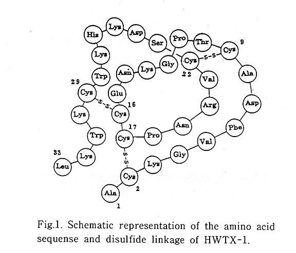
The crude venom from S. huwena was found to be toxic to mice. It consists of 25 compounds with various biological activities and the most abundant component Huwentoxin-1 , abbreviated as HWTX-1 , is found to be a neurotoxin. After purification by reversed phase and ion exchange HPLC its intraperitoneal and intracisternal LD50 in mice are 0.70mg/Kg and 9.40 μg/Kg respectively, causing paralysis of skeletal muscle and rapid respiratory failure. The isoelectric point is 8.95 as determined by the isoelectric focussing electrophoresis technique. It is shown to be a single chain, 33 amino acid residue polypeptide including 6 Cys and 6 Lys and the complete primary sequence as determined by Edman degradation is shown in Fig.1. Assignment of the three disulfide bridges shows that the linkages are formed between the first and fourth Cys, the second and fifth Cys, and the third and sixth Cys, a special pairing pattern seen also in other inhibitor or toxic polypeptides from other biological sources. This was the first spider toxin containing six Cys residues wherein the disulfide bonds have been assigned.
Parallel to these biochemical studies achieved by Liang Songping of the Hunan Normal University, my colleague Zhou Peiai of the National Laboratory of Biomernbrance and Membrance Biotechnology at Peking Unviersity observed, by physiological recordings of the twitch response to electrical stimulations using mouse isolated phrenic nerve diaphragm preparations, that HWTX-1 blocks the neuro-muscular transmission (5). Further experiments proved that HWTX-1 could compete with d- tubocurdrine for binding to post synaptic acetylcholine receptors (6). These results led us to use embryonic Xenopus 'isolated myoballs in culture to study the effect of HWTX-1 on the acetylcholine receptor channel activities with the cell-attached patch clamp technique. Recordings showed obviously that HWTX-1 induces a significant decrease of channel open probability. The mean current amplitude and mean channel open time were also decreased, finally the channel was blocked. These functional studies demonstrated that HWTX-1 is a neurotoxin which binds to the skeletal muscle nACh R.
To probe the structure function relationship ofthis polypeptide I started
to study its solution conformation with the NMR technique. A series of 2D-
COSY, DQF-COSY, TOCSY, and NOESY experiments were performed in
H2O and D2O at pH5.0 and l0-32℃ On Bruker AM-500,
DMX-500, and DMX-600 NMR Spectrometers. All
back bone protons and 98% of the side chain
protons in HWTX-1 were unambiguously assigned.
The three dimensional structures of HWTX-1
were calculated using the program XPLOR 3.1.
The input experimental data included 299
distance constraints (9 disulphide bond constraints
and 290 NOE constraints, including 56 long
range NOEs, 23 medium range NOEs, 82 sequential
NOEs, and 129 intraresidue NOEs), 12φ constraints,
and 4χ1 constraints. 10 best structures
were selected from 43 structures. They all
have NOE violations <0.3A and dihedral violations
<2°. This family of 10 structures has an
average pairwise RMSD of 1.08 (+0.20)Å over
backbone atoms (N, Cα and, C) and 1.91 (± 0.36)Å over all non-hydrogen
heavy atoms (Fig 2). 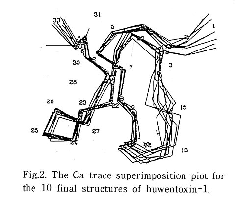
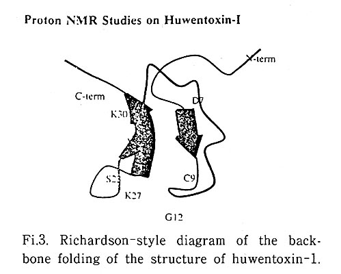
Quality checking of the structure with the PROCHECK program shows that no bad contact exists in the 10 structures and that all residues lie in the most favored regions (80.6% of all residues) or additional allowed regions (the remaining 19.4% of all residues) in the Ramachandran plot. The molecule of HWTX-1 adopts a compact structure consisting of a small triple-stranded anti-parallel β sheet and frve β -turns (Fig3 .) The triple-stranded anti-parallel β-sheet is composed of residues 7-9, 21-23, and 27-30. The frve β-turns involve residues 4-7, 10-13, 11-14, 17-20, and 24-27. The structure of HWTX-1 contains an "inhibitor cystine knot motif" which is composed of a triple-stranded antiparallel β-sheet with three disulphide bonds. This motif is also found in other small polypeptides with diverse biological activities and little amino acid sequence identify from various plants and animals.
Further chemical modification and mutation studies of HWTX-1 are under way in our lab to probe its structure-function relationship and look for possibilities of its application in agricultural or medical uses. Since HWTX-1 competes with d-tubocurarine for binding to nACh R and d-tubo curarine is known to bind its target through a set of two positively charged centers, we make the preliminary proposal that two possible regions in HWTX-1 may be involved in its receptor binding activity: One is around Lys 25 and Lys 27 Iocated in β-turn 24-27; the other is around Ala 1 and Lys 3 near the N-terminals.
The Amaranth α-amylase inhibitor abbreviated as AAI, is a single chain 32 amino acid residues polypeptide isolated from the seeds of the Mexican crop plant Amaranthus hypochondriacus. It is an insect specific α-amylase inhibitor which does not inhibit proteases and mammalian ( including human) α-amylase. It is predicted by my friend Prof. Sandor Pongor at ICGEB, the International Center of Genetic Engineering and Biotechnology in Trieste, Italy to have an inhibitor cystine knot motif. A collaborative project between us, supported by ICGEB, is going on where Dr Pongor has obtained various mutants by engineering the molecule and NMR solution conformation studies are underway in my group.
A new project on molecular bioinformatics supported by the National High Technology and Development Program is added to my group. Through cooperation with the Peking University Computing Center we were approved to be a National Node on the EMBnet (European Molecular Biology Network ) in November, 1996. Our major efforts in 1997 were focussed on establishment of EMBnet data bases on our CERnet (the Chinese Education and Research Net ) to make them easily available to all Chinese scientists. Research on database construction, 3D annotation of new sequences, and development of new algorithms are also started in collaboration with molecular biologists, computer scientists, mathematicians, physicists and chemists both within and outside of Peking Unversity. We are in close contact with scientists working in the fields of human and rice genomics and gene therapy. And we also have collaborative projects on Molecular Bioinformatics with Prof. Edgar Wingender at GBF in Germany and on Advanced Technologies for Protein Purification and Characterisation with Hewlett Packard Laboratories in the US .
LITERATURE: